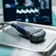
Ultrasound
Gynecological ultrasound — non-invasive diagnostics of uterus, ovaries, and pregnancy with modern ultrasound equipment.
Ultrasound in gynecology
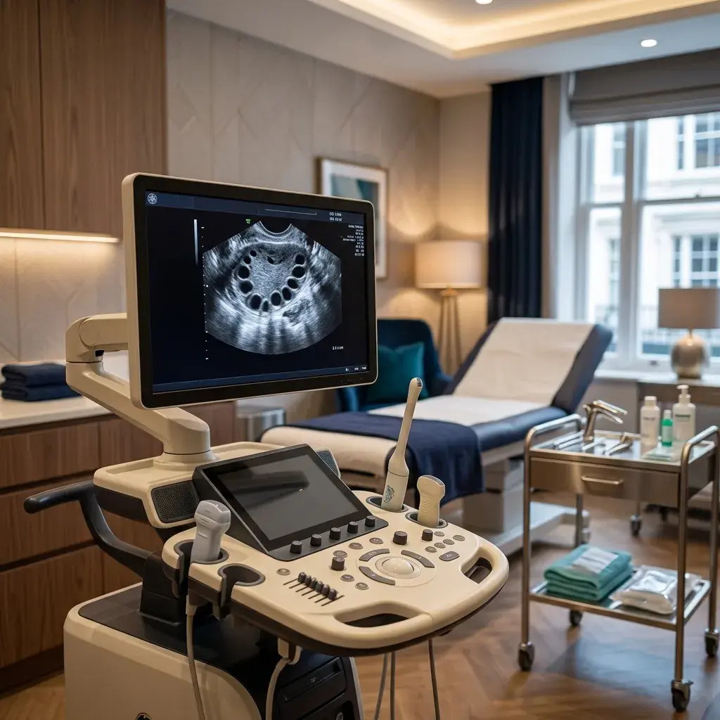
Gynecological ultrasound is a non-invasive diagnostic method using sound waves to visualize the uterus, ovaries, fallopian tubes, and surrounding structures. It is the foundation of modern gynecological diagnostics.
Types of ultrasound examination
- Transvaginal ultrasound — probe inserted vaginally, provides the most detailed pelvic organ imaging
- Transabdominal ultrasound — over the abdomen, used for virgins and during pregnancy
- Power color doppler — assessment of tissue and tumor vascularity
- 3D/4D ultrasound — three-dimensional imaging in pregnancy and anomaly diagnosis
What does gynecological ultrasound detect?
- Ovarian cysts — functional, endometriomas, dermoids
- Uterine fibroids — location, size, and number
- Endometrial polyps — uterine lining thickenings
- Ectopic pregnancy — pregnancy outside the uterus
- Ovulation tracking — follicular monitoring during stimulation
- Congenital anomalies — septum, bicornuate uterus
- Ovarian reserve assessment — antral follicle count (AFC)
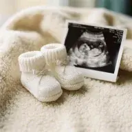
Ultrasound in pregnancy
- Early pregnancy detection — gestational sac visualization from week 5
- Nuchal translucency (NT) — screening for chromosomal anomalies (weeks 11-14)
- Morphology scan — detailed baby anatomy examination (weeks 18-22)
- Fetal vessel doppler — baby's circulation assessment
How to prepare for ultrasound?
For transvaginal ultrasound, no special preparation is needed — the bladder can be empty. For transabdominal, a full bladder is needed (drink 3-4 glasses of water one hour before the exam).

Performed by
Dr Đorđe Petković
Consultant in Operative & Endoscopic Gynaecology · 17+ years of experience
Patients often ask
Yes, ultrasound is completely painless and non-invasive, lasting 10-20 minutes.
Once yearly as part of regular gynecological check-up, or more frequently as recommended.
Ultrasound can identify suspicious changes, but definitive cancer diagnosis requires biopsy with histopathological analysis.