Breast examination
Breast examination — clinical exam, ultrasound, and screening for early detection of changes and breast cancer prevention.
Breast examination — why is it important?
Breast examination is a key part of preventive gynecological check-up helping in early detection of breast tissue changes. Breast cancer is the most common malignancy in women, and early diagnosis significantly improves prognosis and cure chances.
Breast examination methods
- Clinical breast exam — systematic palpation by a gynecologist for lumps, skin dimpling, or nipple changes
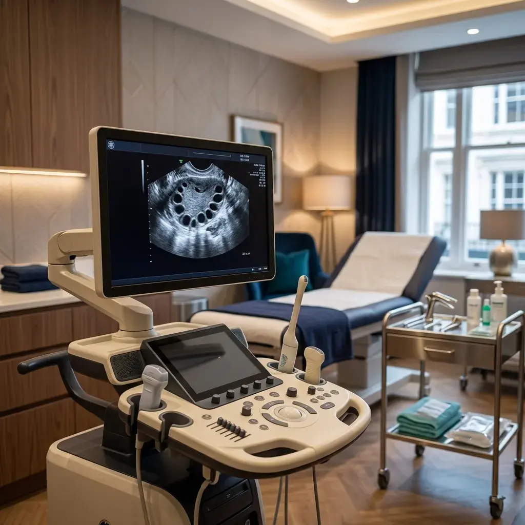
- Breast ultrasound — non-invasive visualization of breast tissue structures, especially useful for young women with dense tissue
- Mammography — X-ray examination, gold screening standard from age 40
- Breast MRI — for high-risk patients or unclear findings
- Biopsy — tissue sampling for histopathological analysis of suspicious lesions
What can examination reveal?
- Fibroadenoma — benign firm lump, most common in young women
- Fibrocystic changes — painful fluid-filled cysts, benign condition
- Papilloma — benign growth in milk duct
- Lipoma — benign fatty growth
- Breast cancer — early detection key to successful treatment
Breast self-examination
Every woman from age 20 should regularly perform monthly breast self-examination (best 7-10 days after period start). Pay attention to: lumps, skin color changes, nipple retraction, nipple discharge.
When to see a gynecologist?
- New lump or thickening found in breast
- Change in breast size, shape, or contour
- Nipple discharge (especially bloody)
- Skin or nipple retraction
- Redness, peeling, or thickening of breast skin

Performed by
Dr Slobodanka Petković
Specialist in Gynaecology & Obstetrics · 35+ years of experience
Patients often ask
Mammography is recommended from age 40 as regular screening every 1-2 years. Women with family history from earlier.
No, these are complementary methods. Ultrasound is better for young women with dense tissue, mammography for detecting calcifications.